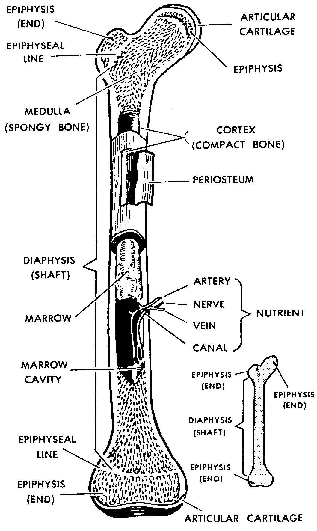
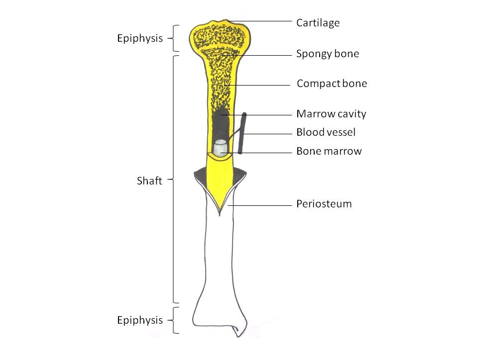
Long Bone Diagram Labeled : 1: Schematic drawing of a longitudinal section through a ... : Each end bone is called an epiphysis (epi = on;
byAdmin•
0
Long Bone Diagram Labeled : 1: Schematic drawing of a longitudinal section through a ... : Each end bone is called an epiphysis (epi = on;. The ilium is the big bone of the hip, the ischium is the bone on which one sits and the pubis forms the lower frontal hip bone as seen in the diagram. This gives you the opportunity to get a general feel of the appearance of each structure and their relations to the structures around them. Physis = to grow) while the middle bone is called a diaphysis (dia = passing through). Jul 29, 2021 · labeled diagram the best way to kick off your revision is with a urinary system diagram which clearly shows all of the structures found within. The longest and the strongest bone in the human skeletal system as you can observe in the labeled skeleton diagram of the human body.
The femur is a type of long bone located in the thigh and is the largest bone of the skeletal system. Download pdf worksheet (blank) download pdf worksheet (labeled) Above, you can see a labeled diagram showing the main bones of the body. They are hard, rigid, and are made up of calcium and phosphorous. Joints hold your bones together and allow your rigid skeleton to move.
Images 04. Skeletal System | Basic Human Anatomy from brooksidepress.org Download pdf worksheet (blank) download pdf worksheet (labeled) Take a look at the urinary system diagram labeled below. Labeled diagram of plant cell, created with biorender.com. They are hard, rigid, and are made up of calcium and phosphorous. May 31, 2021 · now that you know a little bit more about the types and locations of bones, why not test your memory with a bone labeling exercise? Mar 29, 2021 · femur bone anatomy. Joints hold your bones together and allow your rigid skeleton to move. Within the long bones, the epiphysis is the first to undergo conversion followed by the diaphysis before extending to the metadiaphysis 5,6.
The typical characteristics that define the plant cell include cellulose, hemicellulose and pectin, plastids which play a major role in photosynthesis and storage of starch, large vacuoles responsible for regulating the cell turgor pressure.
Within the long bones, the epiphysis is the first to undergo conversion followed by the diaphysis before extending to the metadiaphysis 5,6. Jul 29, 2021 · labeled diagram the best way to kick off your revision is with a urinary system diagram which clearly shows all of the structures found within. Joints hold your bones together and allow your rigid skeleton to move. The typical characteristics that define the plant cell include cellulose, hemicellulose and pectin, plastids which play a major role in photosynthesis and storage of starch, large vacuoles responsible for regulating the cell turgor pressure. The femur is a type of long bone located in the thigh and is the largest bone of the skeletal system. At birth, each long bone is made of three individual bones separated by hyaline cartilage. This gives you the opportunity to get a general feel of the appearance of each structure and their relations to the structures around them. Physis = to grow) while the middle bone is called a diaphysis (dia = passing through). It is attached to the tibia at both the ends. The following diagram and paragraphs explain the skeletal anatomy of a dog. Its upper end articulates with the tibia at the back of its head, whereas while attaching to the tibia with its lower end, it angles slightly forward. Feb 12, 2004 · all of your bones, except for one (the hyoid bone in your neck), form a joint with another bone. The longest and the strongest bone in the human skeletal system as you can observe in the labeled skeleton diagram of the human body.
It is a long bone structure that encases the brain, and contains a cavity called the orbit, where the eye is located. They are hard, rigid, and are made up of calcium and phosphorous. Its upper end articulates with the tibia at the back of its head, whereas while attaching to the tibia with its lower end, it angles slightly forward. This gives you the opportunity to get a general feel of the appearance of each structure and their relations to the structures around them. The anatomy of the femur can be divided into proximal, central, distal, and posterior parts.
Human Anatomy Body - Page 2 of 160 - Human Anatomy for ... from www.anatomylibrary99.com Each end bone is called an epiphysis (epi = on; The typical characteristics that define the plant cell include cellulose, hemicellulose and pectin, plastids which play a major role in photosynthesis and storage of starch, large vacuoles responsible for regulating the cell turgor pressure. Mar 29, 2021 · femur bone anatomy. The fibula is a long but thin bone which, along with the tibia, forms the lower part of the human leg. The ilium is the big bone of the hip, the ischium is the bone on which one sits and the pubis forms the lower frontal hip bone as seen in the diagram. The following diagram and paragraphs explain the skeletal anatomy of a dog. Feb 12, 2004 · all of your bones, except for one (the hyoid bone in your neck), form a joint with another bone. Labeled diagram of plant cell, created with biorender.com.
Above, you can see a labeled diagram showing the main bones of the body.
The femur is a type of long bone located in the thigh and is the largest bone of the skeletal system. Download pdf worksheet (blank) download pdf worksheet (labeled) It is attached to the tibia at both the ends. May 31, 2021 · now that you know a little bit more about the types and locations of bones, why not test your memory with a bone labeling exercise? Jul 29, 2020 · the long bones of the body contain many distinct regions due to the way in which they develop. It is a long bone structure that encases the brain, and contains a cavity called the orbit, where the eye is located. The femur and/or hip may fracture secondary to trauma, so understanding the femur bone anatomy is important. Jul 29, 2021 · labeled diagram the best way to kick off your revision is with a urinary system diagram which clearly shows all of the structures found within. Mar 29, 2021 · femur bone anatomy. Labeled diagram of plant cell, created with biorender.com. Above, you can see a labeled diagram showing the main bones of the body. The femur or the thigh bone is closest to the body. The following diagram and paragraphs explain the skeletal anatomy of a dog.
Take a look at the urinary system diagram labeled below. One extremely important part of a dog's skeletal anatomy is the skull. Also, islands of red marrow may be seen anywhere in the skeleton, typically in a subcortical distribution, often with central yellow marrow giving it a bull's eye appearance on axial imaging. The typical characteristics that define the plant cell include cellulose, hemicellulose and pectin, plastids which play a major role in photosynthesis and storage of starch, large vacuoles responsible for regulating the cell turgor pressure. The fibula is a long but thin bone which, along with the tibia, forms the lower part of the human leg.
Label Ideas 2020: 31 Label The Parts Of A Long Bone from www.jobilize.com Jul 22, 2021 · figure: The femur or the thigh bone is closest to the body. Take a look at the urinary system diagram labeled below. Labeled diagram of plant cell, created with biorender.com. Feb 12, 2004 · all of your bones, except for one (the hyoid bone in your neck), form a joint with another bone. Joints hold your bones together and allow your rigid skeleton to move. Physis = to grow) while the middle bone is called a diaphysis (dia = passing through). Each end bone is called an epiphysis (epi = on;
They are hard, rigid, and are made up of calcium and phosphorous.
One extremely important part of a dog's skeletal anatomy is the skull. It is a long bone structure that encases the brain, and contains a cavity called the orbit, where the eye is located. Its upper end articulates with the tibia at the back of its head, whereas while attaching to the tibia with its lower end, it angles slightly forward. Jul 29, 2021 · labeled diagram the best way to kick off your revision is with a urinary system diagram which clearly shows all of the structures found within. Feb 12, 2004 · all of your bones, except for one (the hyoid bone in your neck), form a joint with another bone. Above, you can see a labeled diagram showing the main bones of the body. Within the long bones, the epiphysis is the first to undergo conversion followed by the diaphysis before extending to the metadiaphysis 5,6. Physis = to grow) while the middle bone is called a diaphysis (dia = passing through). The fibula is a long but thin bone which, along with the tibia, forms the lower part of the human leg. Jul 22, 2021 · figure: The following diagram and paragraphs explain the skeletal anatomy of a dog. At birth, each long bone is made of three individual bones separated by hyaline cartilage. The femur is a type of long bone located in the thigh and is the largest bone of the skeletal system.
The typical characteristics that define the plant cell include cellulose, hemicellulose and pectin, plastids which play a major role in photosynthesis and storage of starch, large vacuoles responsible for regulating the cell turgor pressure long bone diagram. Jul 22, 2021 · figure: